_191119033000.jpg)
Introduction
Low level radioactive waste (LLRW) management is an important element of all radioactive material use programs. If that program generates wastes that must be removed from your site for disposal, LLRW management will consume significant financial resources as well. As the RSO. you want to use your resources as efficiently as possible. Using the information presented here should make efficiency a reality.
Waste Minimization Plan
The most subtle element of any low-level radioactive waste management program is a waste minimization plan. In its simplest form, the plan needs to convey the notion that your institution’s policy is to reduce, where and when possible, the volume of radioactive waste produced. This statement is perhaps obvious, but it needs to be on paper. The details of a conversation you have with a radioactive material user who promises to greatly reduce the magnitude of a waste stream is easily forgotten. A document, however, distributed to all users that clearly states your policy, cannot be ignored without consequences. The minimization plan should tell users, and remind those who approve purchases, that radioactive material acquisitions should be minimized to the quantity needed. The RSO should review all purchases of radioactive material, and give approval before they can be ordered. Avoid volume discounts for the sake of the discount. Also, the plan should state that non-radioactive methods for completing the task at hand should be evaluated and that reusable laboratory items need to replace disposable supplies. Whenever possible, sealed sources should be exchanged with the vendor when new sealed sources are acquired.
This policy is also a good place to list wastes your institution is not equipped to handle and therefore, does not wish generated. The list does not have to be specific (e.g., histology staining solutions containing uranyl acetate and formalin greater than 5% by volume), but should state categories of waste for which disposal is non-existent or prohibitively expensive. Two examples of such mixed wastes would be 1) combinations of radioactive materials and hazardous chemicals, and 2) radioactive materials and heavy metals. There are others. The creation of a list does not have to mean a strict prohibition. It simply means that processes that produce wastes on the “no-no list” should be subjected to a more thorough review. Invite the principal investigators involved with generating the waste to suggest possible solutions. At worst, you have an opportunity to educate the principal investigators as to the problems faced in disposing of some waste forms. At best, those most closely involved with the procedure may suggest a process change or addition that can eliminate or reduce the waste in question. Finally, tap the talent that exists at your institution to solve this problem. The experience and knowledge that a researcher, engineer, or technician has with a family of compounds could provide enough insight to solve the problem. All this effort has another important benefit for you and the radiation safety program. You are seen as a problem solver or a facilitator, not simply a roadblock.
Setting waste minimization goals for each laboratory would also be a good idea, but so far we have not had the time to work with the authorized users to develop and implement such goals. Finally, have the waste minimization plan endorsed by appropriate authorities at your institution. This may mean review by the radiation safety committee, safety committee, CEO, dean, provost, or president. The policy needs some teeth. So unless you are higher in the organization than most RSOs, get the review. The review also is a means of educating senior management about the needs of your radiation protection program. Such an opportunity should not be missed. Your efforts will be worth it because the headaches you avoid will be your own.
Solving Today’s Problems
Now that we have taken care of some of tomorrow’s headaches, how do you handle the ones you have today? The answer to that question depends on three properties of the waste: physical form, half-life, and volume.
Physical Form
Physical form includes the obvious as well as the subtle. The obvious includes solids, liquids and gases. Aqueous liquids containing radioactive materials are obviously different from contaminated paper and plastic. The packaging, handling, storage, and disposal method for each will be different. An example of subtle is sharps. Sharps (needles, lancets, pipettes) are solid, yet they possess the ability to puncture the skin. Therefore, definite guidelines exist for their disposal. Specifically, the disposal of sharps requires leak-proof, puncture resistant containers. The use of these containers may conflict with standard procedures for disposal of radioactive materials. For example, the usual colors associated with radioactive material are yellow and magenta. Sharps containers are red. You must adequately label those containers holding radioactive sharps. In order to protect your radiation safety staff and comply with the law, you need to incorporate the guidelines for packaging into your procedures.
Half- life
Half-life determines whether or not you can consider the waste for decay-in-storage (DIS). Most radioactive material licenses allow certain radioactive waste to be held for ten half-lives and then be disposed of as nonradioactive if no detectable activity remains. The maximum half life is specified in the license. A typical value today is 90 days. If the waste has a half-life less than or equal to 90 days, you may hold the waste for decay. If the half life is over this value, shipment for disposal, and the associated cost, are your only options. The 90-day mark is typical, but not magic. If you use a particular isotope routinely and that isotope has a longer half life (e.g., Sn·113, t1/2 = 115 d), check with your licensing agency. Depending on the circumstances, they may amend your license and allow you to hold that isotope for decay.
Volume
Volume dictates the amount of effort you spend with a particular waste category. For example, if you produce enough paper and plastic laboratory waste contaminated with S-35 (t1/2 = 87 d) to fill one 55-gallon drum (7.5 ft3) each week, you will produce 52 drums each year. To ship that waste at current rates would cost over $80,000. The potential savings make DIS attractive. However, before the first drum is removed from your DIS program, you will accumulate 124 drums (930 ft’) of waste as you wait the required ten half-lives. Your institution may have to commit a portion of its physical plant (space) to handle this aspect of your DIS program. This may mean construction of a storage building or conversion of office, laboratory or production space. A small office (10′ 8″ x 10′ 8″ x 8′) runs about 900 ft’. In actually, you will need more space than just the anticipated volume of waste because not all the space will be able to be utilized. You will have to periodically inspect the drums, and retrieve them for survey after decay. For example, the Albany Medical Center (AMC) built a 15′ x 45′ x 10′ DIS facility for a cost of approximately $50,000. Decaying waste in storage instead of shipping it for burial saved us $43,000 in 1995 and $31,000 in 1996. Alternatively, you could invest in a suitable compactor to help minimize waste volume, but remember the unit itself will require space in which to operate, and will need to be surveyed (and possibly decontaminated) periodically. This example shows that even a straight forward waste issue requires some forethought before the optimal solution is found.
Low-level Radioactive Waste Management at Albany Medical Center
A review of the procedures employed at AMC will illustrate how these concepts are applied. AMC is composed of the Albany Medical Center Hospital and the Albany Medical College. The hospital provides a full range of medical care including clinical laboratories, nuclear medicine, and radiation therapy programs. The medical college, in addition to training physicians, conducts biomedical research using radioactive materials. Therefore, the types of low level radioactive waste generated are typical of that found in both clinical and academic settings.
The first waste group (Category 1) is contaminated with radioisotopes with a half-life of less than four days. The nuclear medicine isotopes technetium- 99m (6.02 h), gallium-67 (3.26 d), and thallium·201 (3.03 d) dominate here. The material is collected from the nuclear medicine clinic in containers suitable for storage including cardboard boxes lined with a plastic bag for dry wastes, appropriately labeled sharps containers for syringes, and plastic bags for various liners and rags. The waste is assigned an inventory number and placed in storage for 10 half-lives (about 30 days). After decay, the waste is surveyed using a count rate meter and scintillation probe. If the readings are indistinguishable from background levels, radioactive labels on containers are defaced or removed, the change in inventory is logged, the waste repackaged and shipped for disposal as regulated medical waste (RMW), a New York State-defined category (10 NYCRR 70). Our efforts focus on the radiation hazard because the components of the waste are well defined with respect to radioactivity and physical form, and the bio-hazard is small.
Another means by which short lived isotopes enter the waste stream subset of this category is directly as regulated medical waste contaminated with radioactive materials. This waste is generated in the hospital, usually from patients who recently underwent a nuclear medicine procedure. Often an item as simple as the gauze used to cover the injection sight retains enough radioactivity to alarm portal monitors used to screen medical waste. This waste is treated in a similar fashion to other nuclear medicine type wastes collected, with one exception. Unlike the waste from the nuclear medicine clinic, this radioactive material is commingled with RMW from general hospital operations. The bio-hazard potential is greater and the probability that it contains material that will putrefy is high, so the material is held for DIS in a freezer to minimize those problems.
The next grouping (Category 2) consists of wastes contaminated by radioisotopes with half-lives between 4 and 30 days. Typical isotopes include iodine-131 (8 d), phosphorus·32 (14.3 d), and rubidium-86 (18.6 d). The I-131 waste results from the hospitalization and treatment of individuals for various thyroid conditions. The waste contains the usual paraphernalia associated with clinical care, including food scraps from meals consumed within the hospital room. The other isotopes are used in biomedical research and contain disposable laboratory items of paper and plastic. Waste is collected from Laboratories and clinical areas, segregated by half-life, and packaged in containers suitable for transport as RMW. The containers, usually cardboard boxes lined with plastic are also labeled as radioactive material and are placed in our DIS facility. Because the iodine waste contains material (food) that will readily putrefy, this waste is stored for decay in a 12′ x 15′ x 7′ walk-in freezer. After an appropriate decay period, the containers are surveyed, the radioactive material label is removed, the disposal is noted in the DIS inventory log, and the material is shipped as RMW.
As discussed earlier, the subtle differences in physical form significantly impact the way the waste is handled. Although a solid, the waste associated with the patient therapy has a unique characteristic that requires attention. In this case. the putrefaction of the material is addressed by using refrigeration.
The third grouping (Category 3) is waste with a half-life between 31 and 65 days (inclusive). The predominant isotope here is iodine-125 (60. 1 d) resulting from research activities. Because of the volume generated, we use a compactor when placing this waste into 55-gallon drums. The volume reduction achieved with the compactor is not tremendous, about 3:1. but compactor use eases some space constraints. Furthermore, the use of a metal drum provides a substantial container for waste that is held close to two years. When full, an inventory number is assigned, the quantity of activity noted, and the drum is placed in our DIS facility. After ten half-lives, the material is removed from the drum, checked for residual radioactivity, and shipped as RMW. The modest compaction ratio achieved with our compactor strikes a balance between the need to control volume and the need to retrieve materiel for survey when DIS is complete.
 The fourth waste grouping (Category 4) is composed of radioisotopes with a half-life between 66 and 90 days. When access to disposal facilities was intermittent, we amended our radioactive material license to allow storage for decay for these longer-lived radioisotopes. The predominant player here is sulfur-35 (87.2 d). Again, the compactor and metal drums are used to help control volume and provide an adequate storage container for the waste. Inventory and removal procedures are Identical with those of the previous group.
Finally, the fifth form of dry, active waste (Category 5) includes those isotopes with a half-life greater than 90 days such as hydrogen-3 (tritium, 12.3 y). carbon-14 (5,730 y). and occasionally calcium-45 (162.7 d). Waste is compacted into drums and periodically shipped for disposal during the year. To prevent confusion with waste held for decay, a separate log is maintained for this long-lived material.
To the greatest extent practical. waste is stored in its ultimate disposal container. Most of the DIS waste is placed in containers suitable for RMW transport and disposal. All the drums used at AMC meet DOT Specification 17H. For long-lived wastes, these drums are required for shipment off site. For those used in the DIS program, we avoid having to repackage the waste should long-lived material inadvertently be placed in them.
In order to ensure that waste awaiting compaction is handled according to proper procedures and not commingled, it is sorted into color-coded bins. Categories 3, 4 and 5 waste are each assigned their own color: blue. black and yellow respectively. Since we have only one drum compactor. the bins allow us to “save up” material of each category and eliminate shuffling partially filled drum into and out of the compactor for just a few bags. The waste is moved from its color-coded storage bin to the like-colored drum. It then becomes a simple matter to prevent confusion when packing, emptying, or shipping a drum. For example, only yellow drums, which contain long lived material should ever be shipped for disposal: only blue and black drums should ever be emptied on site (for confirmatory survey and disposal after decay in storage. Confusion with other waste categories has prevented immediate processing of those wastes. Waste collected from the nuclear medicine clinic (Category 1) is inventoried and placed into a storage area separate from the other wastes immediately after collection. Likewise, the wastes from iodine therapies (Category 2) are placed into storage immediately after collection and in a form easily distinguished from the other wastes that share the freezer.
A unique waste form generated at AMC is animal carcasses contaminated with radioactive materials, which also require special handling. We have four options for handling carcasses. First, if the carcass is contaminated with C-14 or H-3 at 0.05 µCi g-1 or less, then the carcass can be disposed of without regard to its radioactivity [10 CFR 20.2005(a)(2)]. This is a specific regulatory exemption for only two isotopes up to a specific concentration. Although it has limited applicability at AMC, do not overlook its possible use. The second is DIS. With a freezer available, we can hold carcasses the required number of half-lives, then dispose of them as RMW. Third, prepare the carcass for disposal by a commercial URW service. This option may mean incineration or shallow land burial. Because of the cost, this option is rarely used.
The fourth option was homegrown and evolved by using the talents available at the Medical Center. Two researchers have developed a process for hydrolysis of animal tissue using a hot alkali solution at pressure. Their method can reduce 100 kg of animal carcasses into two constituent parts: 1) a solution of amino acids, soaps, and simple carbon compounds, and 2) bones. The solution is sampled for radioactivity and disposed of via the sanitary sewer. The bones are dried and currently shipped as regulated medical waste, although if crushed, could provide a source for bone meal. The process has met with regulatory approval for the disposal of radioactive material and potentially infectious materials. It costs about $0.13 to dispose of a kilogram of animal carcass compared to about $85 through other commercial source at depth in tissue as well as room scatter.
Results and Discussion
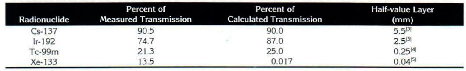
Table 1 shows the transmission and therefore the degree of attenuation provided by the 0.5-mm lead equivalent apron for predominantly primary photons emitted for the various radionuclides, attenuation condition (1). As expected, the apron renders little protection against the high-energy photons from Ir-192 and Cs-137. However, measurable attenuation is noted for these high energy photon emissions in the relatively thin lead-equivalent apron.
The measured transmission values presented in Table 1 differ from values which may be calculated from reported half-value layers for lead at various energies where mono-energetic beams and good geometry are assumed. For Cs-137, Ir-192 and Tc-99m, the measured transmission is less than that predicted by exponential attenuation. The decreased transmission through the lead apron may be due to an inflated value for the initial exposure rate due to scattered radiation created in the concrete block shield. Thus, the efficient attenuation by the lead apron of this lower quality radiation would result in an apparent decreased transmission measurement. With Xe-133, a relatively large measured transmission measurement is noted as compared to the calculated value. Characteristic lead x-rays are efficiently produced in the lead apron due to the incident 81-keV photon energy. These x-rays will be well transmitted through the lead and thus, the x-ray production in the lead apron.
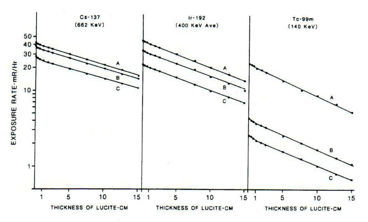
As expected, the data shown in Figure 1 do not yield a single exponential pattern. In all cases, the transmission measurements will be the result of attenuation of both primary and scattered radiation. The three measurement conditions sequentially add additional scatter material resulting in the configuration which best simulates the true clinical condition. Since all three curves for a given radionuclide are essentially the same shape, it is reasonable to assume that it is inconsequential where the scatter originates. Thus, the depth of the radionuclide in Lucite or tissue, the presence of the lead and any other sources of scatter do not alter the quality of the radiation to a significant degree. Further, the presence of the lead apron reduced the exposure. No enhancement due to scattered radiation in the lead apron was discerned. This can be noted by comparing the initial values of curves A and B for each radionuclide .
Using the Berger equation, for a point isotropic source and an infinite medium, factors were calculated to estimate the dose build-up from scatter created in the lead apron.For the 0.5-mm lead apron, the dose build-up factor is 1.018 for the 662-keV photons of Cs137. In comparison, exponential attenuation predicts a 6% decrease in exposure. Thus, a net reduction should be expected due to the presence of the lead apron. This correlates with the net exposure rate reduction presented in the data.
As the energy of the photon decreases, the build-up factor also decreases. Thus, the 1.8% build-up for Cs-137 should be the maximum build-up factor for the radionuclide investigated. Therefore, the build-up phenomena for Ir-192 and Tc-99m, again, should not be Significant in comparison with attenuation. This Is also in agreement with the data presented.
Conclusion
Based upon the measurements and calculations presented, we offer the following for consideration: with respect to the question of the nursing staff and brachytherapy personnel wearing lead aprons when dealing with Cs-137 and Ir-192, the lead apron affords little reduction in exposure rates, as expected. This slight reduction in exposure rate may not warrant the false sense of security conveyed to these personnel by using the lead aprons. Time, distance, and significant shielding (i.e., bedside shields) are still the overriding factors to consider. The presence of the lead apron, however, should not enhance the radiation exposure to the wearer when exposed to Cs-137 or Ir-192. With regard to radionuclides utilized in nuclear medicine, such as Tc·99m and Xe-133, the lead apron furnishes a significant degree of protection. However, the actual utilization of lead aprons should be evaluated with regard to the “cost-benefit” associated with its use. Using a lead apron may reduce mobility, thereby increasing the time required to perform a particular operation. Still, situations may arise in which one may want to use a lead apron. Such situations involve handling high activities, “milking” generators, pregnancy of technicians, extended dose proximity with uncooperative patients, etc.
by Brian M. Methe for RSO Magazine
References
1. Bushong. Stewart c.. Radiologic Science for Technologists, 3rd Edition, St. Louis: C.V. Mosby. 1984; p. 545.
2. Eberline Model RO-2 Ion Chamber Technical Manual. Eberline Instruments, P.O. Box 2108. Sana Fe, NM 87504-2108.
3. Khan. Fail M .• The Physics of Radiation Therapy, Baltimore: Williams and Wilkins, 1984; p. 355.
4. Radiological Health Handbook. U.S. Department of Health. Education and Welfare, Bureau of Radiological Health, Rockville, MD. 1970; p. 163.
5. Xeneisol Xe-133 package insert R8IB2. Mallinckrodt, Inc .• St. Louis. MO 63134.
6. Schaeffer. N.M., Reactor Shielding for Nuclear Engineers. U.S. Atomic Energy Commission. Oak Ridge. TN. 1973; p. 171 · 172.
The Authors
Howard R. Elson is the Chief Physicist of the Division of Radiation Oncology at the Barrett Center for Cancer Prevention. and Director of Graduate Studies. Treatment and Research at the University of Cincinnati
College of Medicine. He is a qualified radiation expert, medical and non-medical. registered with the state of Ohio. Dr. Elson holds degrees from the University of Chattanooga (BA. physics), Emory University (MS.
physics). and the University of Cincinnati (PhD. biomedical nuclear engineering).
University of Cincinnati Medical Center Division of Radiation Oncology Charles M. Barrett Center P.O. Box 670757 Cincinnati,OH 45267-0757 phone: 513-558-5668
Steven J. Lukes is clinical staff medical physicist at The Christ Hospital in Cincinnati, Ohio. He holds .!I BS in science and an MS in radiologic sciences from the University of Cincinnati. Mr. Lukes is certified by
the American Board of Radiology in therapeutic and medical nuclear physics, and an NMT certification in nuclear medical technology.